432001-19-9
432001-19-9 structure
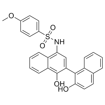
- Name: C188-9
- Chemical Name: KZ3DLD11RQ
- CAS Number: 432001-19-9
- Molecular Formula: C27H21NO5S
- Molecular Weight: 471.524
- Catalog: Signaling Pathways JAK/STAT Signaling STAT
- Create Date: 2018-07-08 11:26:56
- Modify Date: 2026-07-09 12:57:07
-
C188-9 is a Stat3 inhibitor, with a Kd of 4.7 nM.
| Name | KZ3DLD11RQ |
|---|---|
| Synonyms |
N-(1',2-Dihydroxy-1,2'-binaphthalen-4'-yl)-4-methoxybenzenesulfonamide
KZ3DLD11RQ C188-9 |
| Description | C188-9 is a Stat3 inhibitor, with a Kd of 4.7 nM. |
|---|---|
| Related Catalog | |
| Target |
STAT3:4.7 nM (Kd) |
| In Vitro | C188-9 is a Stat3 inhibitor, with a Kd of 4.7 nM[1]. The IC50s of C188-9 to inhibit Stat3 activation in AML cell lines are in the range of 4-7 μM, and in primary AML samples the IC50s are in the range of 8-18 μM. For apoptosis studies, AML cell lines and primary samples are treated for 24 hours with C188-9, then apoptotic cells are quantified by FACS analysis for annexin V-labeled cells. The EC50s for apoptosis induction are quite variable, ranging from 6 μM to over 50 μM[2]. |
| In Vivo | Of the approximately 13,528 discernible genes, levels of 37 gene transcripts are altered by C188 (17 down and 20 up-regulated, fdr <0.01, fold change≥1.5), of which 7 are known STAT3 gene targets. In comparison, C188-9 affects a much greater number of genes involved in oncogenesis (384 total, 95 down- and 289 up-regulated), including 76 genes previously reported as regulated by STAT3 (38 down-regulated and 38 up-regulated). Among the 38 genes previously shown to be upregulated by STAT3, 24 (63%) genes are downregulated by C188-9 treatment, as expected. Additionally, 10 more genes downregulated by C188-9 (fdr <0.01, fold change≥1.5) that previously are shown to be upregulated by STAT1. Thus, 40 of 48 (83.3%) genes downregulated by C188-9 previously are shown to be positively regulated by STAT1, including sixteen genes shown to be co-regulated by STAT3 and STAT1. This analysis raises the possibility that the effect of C188-9 on gene transcript levels in HNSCC tumors is mediated by its effects on both STAT3 and STAT1[3]. |
| Cell Assay | Cell lines are plated at 2 to 5×10⁵ cells/mL in growth medium and treated with increasing doses of inhibitor for 24 hours. CD34+ AML cells are plated at 1 to 2×10⁵ cells/mL in IMDM with 20% FBS and Pen/Strep, and incubated with C188-9 (0.3 to 100 μM) for 48 hours. Cells are then harvested and labeled. The fraction of spontaneous apoptosis is determined from an untreated sample and then subtracted from the drug-treated samples to yield the percentage of apoptosis attributed to drug treatment[1]. |
| Animal Admin | Mice[3] UM-SCC-17B cells (1.5×10⁶) are injected into the tongues of athymic, 8-10 week old, male, nude mice. Once tumors are established, mice (20 total; 10/group) are randomized (average tumor vol ~15-20 mm3) to receive 5 times a week, intraperitoneal injections of either DMSO or C188 (50 mg/kg) or C188-9 (100 mg/kg). Tumor volumes are measured twice weekly. Average tumor volumes 6/πx (long dimension) x (short dimension)2)) are calculated and normalized to the volume at first day of treatment and plotted comparison is done by t test (* p<0.05) [3]. |
| References |
| Density | 1.4±0.1 g/cm3 |
|---|---|
| Boiling Point | 680.9±65.0 °C at 760 mmHg |
| Molecular Formula | C27H21NO5S |
| Molecular Weight | 471.524 |
| Flash Point | 365.6±34.3 °C |
| Exact Mass | 471.114044 |
| LogP | 5.24 |
| Vapour Pressure | 0.0±2.2 mmHg at 25°C |
| Index of Refraction | 1.732 |
| Storage condition | -20℃ |