Tissue characteristics of root resorption areas in transplanted maxillary canines.
T Berglundh, B Thilander, S Sagne
Index: Acta Odontol. Scand. 55(4) , 206-11, (1997)
Full Text: HTML
Abstract
The aim of the present study was to describe some histopathologic features of tissues collected from root resorption areas of maxillary canines after transalveolar transplantation surgery. In 8 of 101 transplanted canines, complications including cervical root resorption occurred between 6 and 11 years after treatment. The resorptive processes were located at the supra-alveolar portions of the distal and/or mesial aspects of the teeth and were scheduled for treatment involving surgical exploration. The resorption cavities, which extended from the cementoenamel junction to a position immediately below the bone crest, were filled with a granulation tissue. In four of the diagnosed complication cases, this granulation tissue was carefully excised concomitant with the adjacent gingival tissue after flap elevation and placed in a buffered fixative. After proper soft-tissue healing, the cavities were filled with a glass-ionomer material. The collected biopsy specimens were, after fixation and, in one case, decalcification in ethylenediaminetetraacetic acid, dehydrated and embedded in Epon. Sections 3 microns thick were produced, stained in periodic acid-Schiff and toluidine blue, and used for histometric and morphometric analyses. The histologic analysis showed that the dissected tissue harbored well-encapsulated areas of inflammatory infiltrates. The lesions comprised a relatively low volume of collagen and a large number of inflammatory cells, predominantly lymphocytes.
Related Compounds
| Structure | Name/CAS No. | Molecular Formula | Articles |
|---|---|---|---|
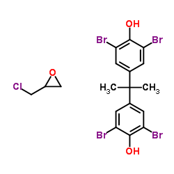 |
Bisphenol A diglycidyl ether, brominated
CAS:40039-93-8 |
C18H17Br4ClO3 |
|
Optimization of a histopathological biomarker for sphingomye...
2012-08-01 [J. Histochem. Cytochem. 60(8) , 620-9, (2012)] |
|
Effect of fixation and embedding on Raman spectroscopic anal...
2006-06-01 [Calcif. Tissue Int. 78(6) , 363-71, (2006)] |
|
Epoxy resin as fixative during freeze-substitution.
2005-11-01 [J. Struct. Biol. 152(2) , 92-103, (2005)] |
|
The role of a new liquid-solid scintillator, a mixture of DP...
1998-07-01 [J. Vet. Med. Sci. 60(7) , 795-7, (1998)] |
|
Platinum blue as an alternative to uranyl acetate for staini...
2007-04-01 [Arch. Histol. Cytol. 70(1) , 43-9, (2007)] |