| Structure | Name/CAS No. | Articles |
|---|---|---|
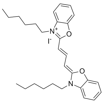 |
3,3'-Dihexyloxacarbocyanine iodide
CAS:53213-82-4 |
|
![N-{6-[(7-nitro-2,1,3-benzoxadiazol-4-yl)amino]hexanoyl}sphingosine Structure](https://image.chemsrc.com/caspic/435/86701-10-2.png) |
N-{6-[(7-nitro-2,1,3-benzoxadiazol-4-yl)amino]hexanoyl}sphingosine
CAS:86701-10-2 |