Inhibition of nitric oxide synthase ameliorates cellular injury in sickle cell mouse kidneys.
N Bank, M Kiroycheva, P C Singhal, G M Anthony, G J Southan, C Szabo
Index: Kidney Int. 58(1) , 82-9, (2000)
Full Text: HTML
Abstract
In previous studies of transgenic sickle cell mice, increased renal expression of inducible nitric oxide synthase (iNOS) and endothelial cell isoform of NOS (EcNOS) was found by Western blot and immunohistochemistry. In addition, putative evidence of peroxynitrite (ONOO-) formation was found in the form of positive immunostaining and immunoblot for nitrotyrosine. Apoptosis was also detected by DNA strand breakage and TUNEL assay. The present study was carried out to examine the role of NO/ONOO- in mediating renal tubular cell apoptosis in sickle cell mouse kidneys.Mercaptoethylguanidine (MEG), a compound that selectively inhibits iNOS and also is a scavenger of ONOO-, was administered intraperitoneally over a five-day period to control and betas mice. Immunohistochemistry of iNOS and nitrotyrosine, DNA electrophoresis, ApoTACS assay for apoptosis, and Western blot of poly(ADP-ribose) polymerase (PARP) were carried out.MEG administration virtually eliminated renal immunostaining of iNOS and nitrotyrosine and prevented DNA strand breakage. In addition, Western blot analysis of PARP, a nuclear DNA-reparative enzyme activated in response to DNA strand breakage, was found to be cleavaged in hypoxic betas mice, but was partially protected in MEG-treated betas hypoxic mice. Finally, apoptosis was markedly reduced by MEG in betas hypoxic mice.These observations provide evidence that NO and/or ONOO- are responsible for initiating cell damage, which leads to apoptosis in sickle cell mouse kidneys.
Related Compounds
| Structure | Name/CAS No. | Molecular Formula | Articles |
|---|---|---|---|
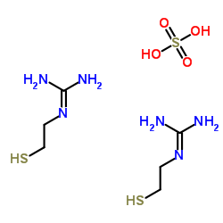 |
MEG hemisulfate
CAS:3979-00-8 |
C6H20N6O4S3 |
|
Nitric oxide is synthesized in acute leukemia cells after ex...
2004-11-08 [Cancer Lett. 215(1) , 43-52, (2004)] |
|
Mitogen-activated protein kinase phosphorylation in kidneys ...
2000-06-01 [J. Am. Soc. Nephrol. 11(6) , 1026-32, (2000)] |
|
Scavenging of peroxynitrite reduces renal ischemia/reperfusi...
2008-01-01 [Ren. Fail. 30(7) , 747-54, (2008)] |
|
Inhibition of experimental gingivitis in beagle dogs with to...
2006-03-01 [J. Periodontol. 77(3) , 385-91, (2006)] |
|
Mercaptoethylguanidine attenuates inflammation in bacterial ...
2000-06-16 [Life Sci. 67(4) , 365-72, (2000)] |