Contrast enhanced CT attenuation correlates with the GAG content of bovine meniscus.
Bejamin A Lakin, Daniel J Grasso, Rachel C Stewart, Jonathan D Freedman, Brian D Snyder, Mark W Grinstaff
Index: J. Orthop. Res. 31(11) , 1765-71, (2013)
Full Text: HTML
Abstract
We determined whether contrast-enhanced computed tomography (CECT) attenuation obtained using a µCT scanner correlated with the glycosaminoglycan (GAG) content and distribution in ex vivo bovine menisci. Bovine samples were immersed in different concentrations of the contrast agents CA4+ and Ioxaglate, and the µCT images were compared to Safranin-O staining. CA4+ and Ioxaglate diffusion-in kinetics and the correlation between their CECT attenuations and GAG content were investigated. CA4+ and Ioxaglate both reached steady state in the meniscal regions within 95 h, with tau values of 20.6 ± 3.98 and 25.9 ± 3.71 h (mean ± SD), respectively. Both agents diffused preferentially through the proximal and secondarily through the distal surface. The CA4+ CECT attenuation was strongly and positively correlated with the GAG content of the meniscus regions (R(2) = 0.89, p < 0.001) at low concentrations (12 mgI/ml), while the Ioxaglate CECT attenuation was moderately and negatively correlated with the GAG content (R(2) = 0.51, p = 0.03) at 60 mgI/ml. CECT can image ex vivo menisci, and the CA4+, compared to Ioxaglate, enhanced attenuation strongly correlates with the GAG content and distribution in bovine meniscus.© 2013 Orthopaedic Research Society. Published by Wiley Periodicals, Inc.
Related Compounds
| Structure | Name/CAS No. | Molecular Formula | Articles |
|---|---|---|---|
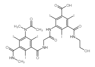 |
ioxaglic acid
CAS:59017-64-0 |
C24H21I6N5O8 |
|
Computed tomography detects changes in contrast agent diffus...
2011-10-01 [Osteoarthr. Cartil. 19(10) , 1190-8, (2011)] |
|
Hyperosmolaric contrast agents in cartilage tomography may e...
2012-02-02 [J. Biomech. 45(3) , 497-503, (2012)] |
|
Acute thyrotoxicosis secondary to destructive thyroiditis as...
2011-04-01 [Thyroid 21(4) , 443-9, (2011)] |
|
The critical role of CT angiography in the detection and the...
2013-01-01 [JBR-BTR 96(2) , 78-80, (2013)] |
|
A rare case of acute contrast-induced sialadenitis after per...
2016-02-01 [Isr. Med. Assoc. J. 15(10) , 652-3, (2013)] |