| Structure | Name/CAS No. | Articles |
|---|---|---|
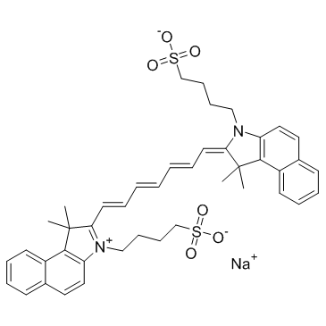 |
Indocyanine Green
CAS:3599-32-4 |
|
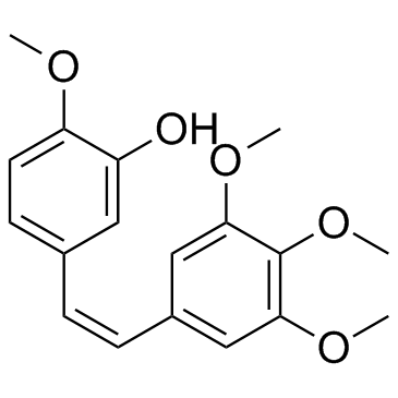 |
Combretastatin A4
CAS:117048-59-6 |