Rinsho byori. The Japanese journal of clinical pathology 2011-09-01

[Histochemical staining methods for lanthanum].

Makoto Miyagawa

Index: Rinsho Byori. 59(9) , 844-51, (2011)

Full Text: HTML

Abstract

In recent years lanthanum compounds have been widely used in the optical and electronic industries. Although release of lanthanum (La) into the environment and exposure to humans are feared, acute or chronic biologic effects of La remain to be elucidated. The present study was undertaken to establish the experimental animal model for La toxicity and histological staining methods for La. After intraperitoneal injections of lanthanum chloride, white precipitates deposited on the surface of the liver. Existence of La in the precipitates was confirmed by a X-ray fluorescent microanalysis. Liver tissues from La treated rats were fixed in paraformaldehyde, embedded in paraffin and cut into thin sections which were used as a La containing standard section. Several reagents for histological stains and spectrophotometry for metals were applied in both test-tube experiments and stainings of tissue sections to test for La. Alizarin complexone (ALC) was found of capable of staining La in tissue. A simple new technique used was described for light microscopic detection of La.

Related Compounds

Structure Name/CAS No. Articles
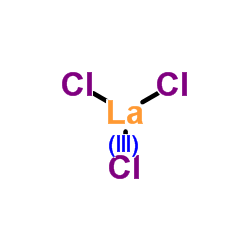
Lanthanum(III) chloride Structure Lanthanum(III) chloride
CAS:10099-58-8
alizarin complexone Structure alizarin complexone
CAS:3952-78-1
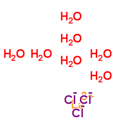
lanthanum chloride heptahydrate Structure lanthanum chloride heptahydrate
CAS:10025-84-0